Breakthrough Study Reveals How GPCRs Dynamically Recognize and Activate G Proteins
New Insights Into a Fundamental Cellular Signaling System
A major advance in molecular biology has shed light on how G-protein-coupled receptors (GPCRs) dynamically recognize and activate heterotrimeric G proteins—an essential process underlying countless physiological responses in the human body. Using high-resolution cryo-electron microscopy, researchers have mapped the structural interactions between the human neurotensin receptor type 1 (NTSR1) and multiple G-protein subtypes, revealing a level of flexibility and adaptability that reshapes current understanding of cellular signaling.
GPCRs form one of the largest and most important families of membrane proteins in humans. They act as molecular sensors, detecting signals such as hormones, neurotransmitters, and environmental stimuli, then translating them into intracellular responses. These receptors are also the target of a substantial proportion of modern pharmaceuticals, making insights into their function both scientifically and economically significant.
The new findings highlight that GPCRs are not rigid, one-size-fits-all structures. Instead, they possess a dynamic intracellular surface capable of reorganizing itself to interact with different G-protein subtypes, a feature that may explain their versatility and efficiency across diverse signaling pathways.
Cryo-EM Unlocks Structural Complexity
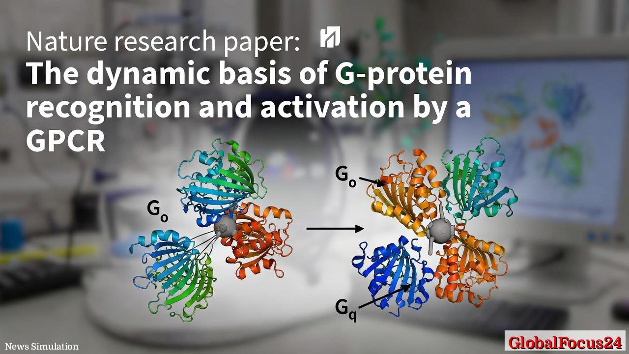
Cryo-electron microscopy (cryo-EM), a technique that allows scientists to visualize biomolecules at near-atomic resolution without crystallization, played a central role in the discovery. By capturing multiple structural states of NTSR1 bound to minimally modified G_o and G_q proteins, researchers observed how the receptor adapts its conformation depending on the G protein it engages.
These structures revealed that the receptor’s intracellular domain undergoes subtle but crucial rearrangements. Rather than adopting entirely distinct conformations for each G protein, the receptor uses a shared set of structural adjustments that can accommodate different partners. This “modular flexibility” suggests a conserved mechanism that balances specificity with efficiency.
The ability to observe these interactions in multiple states marks a significant improvement over earlier structural studies, which often relied on static snapshots. The dynamic view provided by cryo-EM offers a more realistic representation of how these molecular machines function in living cells.
Dynamic Rearrangement Enables G-Protein Recognition
At the heart of the discovery is the finding that GPCRs employ common intracellular rearrangements to recognize different G-protein subtypes. These rearrangements occur primarily in the receptor’s cytoplasmic regions, where G proteins bind.
Key structural elements shift position to create a binding interface that can accommodate variations in G-protein structure. This adaptability allows a single receptor to interact with multiple G proteins, a property known as “coupling promiscuity.”
At the same time, the receptor maintains the ability to selectively activate a specific G protein once binding occurs. This dual capability—broad recognition combined with precise activation—has long puzzled researchers and is now better understood through these structural insights.
Stepwise Activation and Gβγ Separation
The study also provides a detailed look at how G proteins become activated after binding to the receptor. Activation involves the separation of the Gα subunit from the Gβγ dimer, a critical step that enables downstream signaling.
Researchers found that this separation occurs through a stepwise remodeling of specific regions within the Gα subunit, known as switches I, II, and III. These structural elements undergo coordinated changes that weaken the interaction between Gα and Gβγ, eventually leading to their dissociation.
This stepwise mechanism contrasts with earlier models that suggested a more abrupt activation process. The new findings indicate that activation is a gradual, multi-stage event, offering multiple points at which cellular regulation can occur.
Such granularity could prove valuable for drug development, as targeting specific stages of activation may allow for more precise modulation of signaling pathways.
Distinct Dissociation Pathways for G Proteins
Another key discovery involves the way different G proteins disengage from the receptor after activation. The study shows that G_i proteins follow a dissociation pathway that differs significantly from that of G_s proteins.
In particular, the researchers observed divergence between canonical and non-canonical receptor–G_i complexes. These complexes not only differ in their initial binding configurations but also follow separate trajectories as they dissociate from the receptor.
This finding suggests that the signaling outcome may depend not only on which G protein is activated but also on how it disengages from the receptor. Such differences could influence the duration and intensity of the cellular response.
Understanding these pathways in detail may help explain why certain drugs produce varying effects even when targeting the same receptor, a phenomenon known as functional selectivity or biased signaling.
Historical Context of GPCR Research
The study builds on decades of research into GPCR biology, a field that has evolved dramatically since the first receptor structures were solved in the early 2000s. Early work focused on identifying receptor sequences and understanding their basic function, while later studies used X-ray crystallography to reveal static structural snapshots.
The introduction of cryo-EM has transformed the field by enabling researchers to capture multiple conformational states and transient interactions. This shift from static to dynamic structural biology has opened new avenues for understanding how GPCRs operate in real time.
Over the past two decades, GPCR research has been recognized with multiple Nobel Prizes, reflecting its importance in both basic science and medicine. The latest findings represent another step forward, moving from structural description to mechanistic understanding.
Economic and Pharmaceutical Implications
GPCRs are estimated to be the target of roughly one-third of all approved drugs, spanning treatments for cardiovascular disease, neurological disorders, metabolic conditions, and more. As a result, any advance in understanding GPCR function carries significant economic implications.
The ability to map how receptors interact with different G proteins could lead to the development of more selective drugs with fewer side effects. For example, a drug designed to stabilize a specific receptor conformation might preferentially activate one signaling pathway while avoiding others.
This precision could improve therapeutic outcomes and reduce adverse reactions, potentially lowering healthcare costs associated with drug-related complications. It could also accelerate drug discovery by providing clearer structural targets for pharmaceutical design.
Biotechnology firms and major pharmaceutical companies are already investing heavily in structure-based drug design, and findings like these are likely to influence future research pipelines.
Regional Research Trends and Global Collaboration
The advancement underscores the global nature of modern biomedical research. Cryo-EM facilities, which are expensive and technically demanding, are concentrated in major research hubs across North America, Europe, and East Asia.
In the United States, institutions in regions such as California and Massachusetts have been at the forefront of GPCR research, supported by strong funding from both public agencies and private industry. Europe has contributed significantly through collaborative research networks, while countries like Japan and China have rapidly expanded their cryo-EM capabilities in recent years.
This international landscape has fostered a competitive yet collaborative environment, accelerating the pace of discovery. Shared data repositories and cross-border partnerships have become increasingly common, allowing researchers to build on each other’s findings.
Broader Impact on Biomedical Science
Beyond GPCRs, the insights gained from this study may influence the broader field of protein dynamics. The concept that proteins can use shared structural rearrangements to interact with multiple partners could apply to other signaling systems as well.
This perspective challenges traditional views of protein function as rigid and highly specific, instead emphasizing flexibility and adaptability. Such a shift has implications for how scientists approach everything from enzyme design to synthetic biology.
The study also highlights the importance of capturing transient states in biological systems. Many critical processes occur on timescales and in configurations that are difficult to observe, but advances in imaging technology are beginning to bridge that gap.
Future Directions in GPCR Research
Researchers are expected to build on these findings by exploring additional GPCR–G protein combinations and investigating how other factors, such as ligands and membrane environment, influence receptor behavior.
Key questions remain, including how different ligands bias receptor conformation and how intracellular signaling networks integrate multiple inputs. Further studies may also examine how mutations in GPCRs contribute to disease by altering their dynamic properties.
As the field moves forward, the integration of structural biology, computational modeling, and cellular assays will likely play a central role in unraveling the complexity of GPCR signaling.
The latest discovery marks a significant step toward a more complete understanding of one of the body’s most essential communication systems, with implications that extend from fundamental biology to the future of medicine.